Imaging Modalities in Ocular Oncology
Expert-defined terms from the Postgraduate Certificate in Ocular Oncology course at LearnUNI. Free to read, free to share, paired with a professional course.
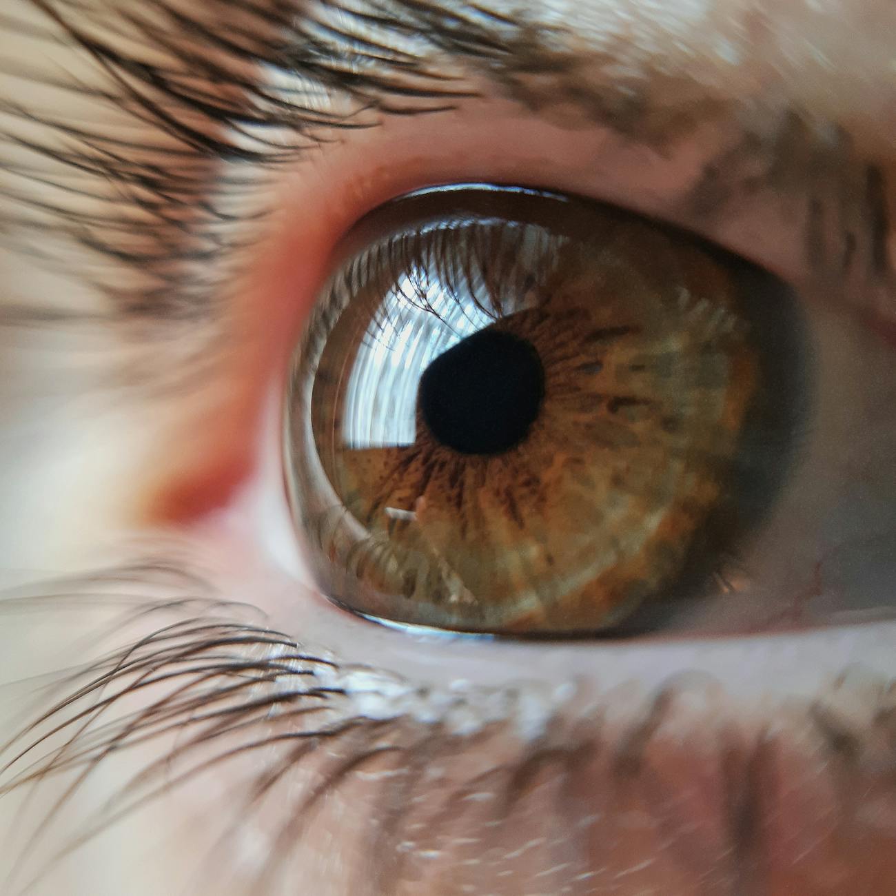
Imaging Modalities in Ocular Oncology #
Imaging Modalities in Ocular Oncology
Imaging modalities play a crucial role in the diagnosis, management, and monitor… #
Various imaging techniques are utilized to visualize and characterize tumors in different parts of the eye. The choice of imaging modality depends on the location, size, type, and extent of the tumor. In ocular oncology, the most commonly used imaging modalities include ultrasound, optical coherence tomography (OCT), fluorescein angiography (FA), indocyanine green angiography (ICGA), fundus photography, and magnetic resonance imaging (MRI).
Ultrasound (US) #
Ultrasound (US)
Ultrasound is a non #
invasive imaging technique that uses high-frequency sound waves to visualize intraocular structures. In ocular oncology, ultrasound is particularly useful for evaluating tumors in the posterior segment of the eye, such as choroidal melanoma. A-scan ultrasound provides information about tumor size and internal reflectivity, while B-scan ultrasound allows for the visualization of tumor shape, location, and relationship to surrounding structures.
Optical Coherence Tomography (OCT) #
Optical Coherence Tomography (OCT)
OCT is a non #
invasive imaging technique that uses light waves to create cross-sectional images of the retina and choroid. In ocular oncology, OCT is valuable for assessing tumor thickness, presence of subretinal fluid, and associated retinal changes. OCT angiography can also provide information about tumor vascularity and neovascularization.
Fluorescein Angiography (FA) #
Fluorescein Angiography (FA)
FA is a diagnostic imaging technique that involves the intravenous injection of… #
In ocular oncology, FA can help identify tumor vascularity, leakage, and associated retinal changes. Late-phase FA may reveal tumor staining and enhance the delineation of tumor margins.
Indocyanine Green Angiography (ICGA) #
Indocyanine Green Angiography (ICGA)
ICGA is an imaging modality that utilizes indocyanine green dye to visualize the… #
In ocular oncology, ICGA can be useful for detecting choroidal tumors, such as choroidal hemangioma. It provides information about choroidal circulation, areas of hyperpermeability, and the presence of polypoidal choroidal vasculopathy.
Fundus Photography #
Fundus Photography
Fundus photography involves capturing detailed images of the retina, optic nerve… #
In ocular oncology, fundus photography is essential for documenting tumor characteristics, such as color, shape, size, and location. It is also valuable for monitoring tumor growth over time.
Magnetic Resonance Imaging (MRI) #
Magnetic Resonance Imaging (MRI)
MRI is a non #
invasive imaging modality that uses magnetic fields and radio waves to generate detailed images of the eye and orbit. In ocular oncology, MRI can be helpful for evaluating intraocular and orbital tumors, such as optic nerve glioma. Gadolinium contrast-enhanced MRI can provide information about tumor vascularity and extension into adjacent structures.
In addition to these imaging modalities, newer techniques such as positron emiss… #
The integration of multiple imaging modalities allows for a comprehensive evaluation of ocular tumors and facilitates treatment decision-making.
Overall, imaging modalities are essential tools in the management of ocular onco… #
Radiologists, ophthalmologists, and oncologists work together to interpret imaging findings and develop individualized treatment plans for patients with ocular tumors.