
Preservation Techniques
Formaldehyde is the cornerstone chemical in traditional embalming, acting as a powerful protein cross‑linker that stabilises tissue structure. It penetrates rapidly through vascular channels, reacting with amino groups to form methylene bri…
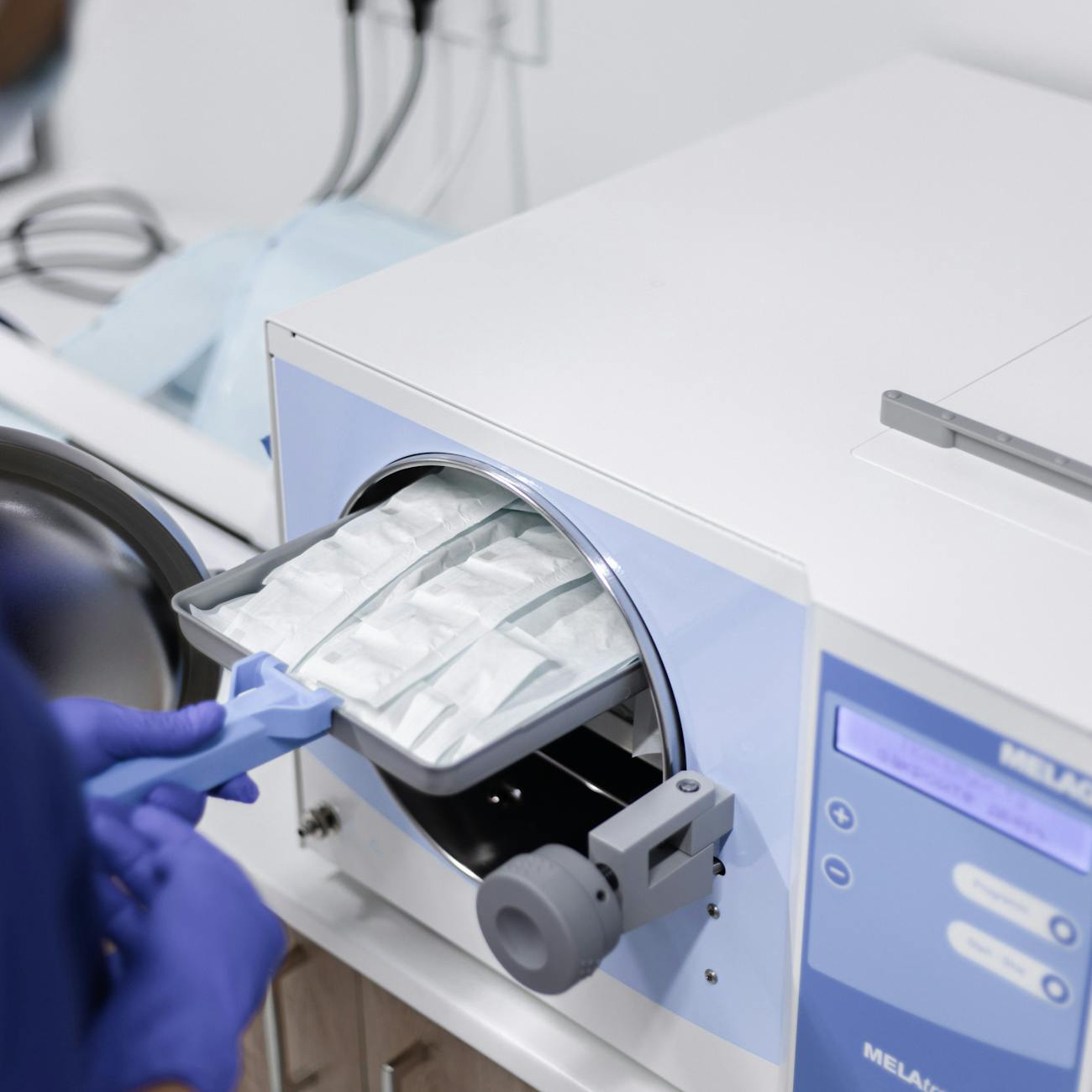
Formaldehyde is the cornerstone chemical in traditional embalming, acting as a powerful protein cross‑linker that stabilises tissue structure. It penetrates rapidly through vascular channels, reacting with amino groups to form methylene bridges, which halt enzymatic breakdown and bacterial growth. The typical concentration in arterial fluid ranges from 3 % to 5 % w/v, often buffered with sodium bicarbonate to maintain a pH between 6.8 And 7.2. In practice, an embalmer may prepare a mixture containing 4 % formaldehyde, 0.5 % Phenol, and a humectant such as glycerol, then inject it under a pressure of 150 mm Hg to ensure uniform distribution. Challenges arise from formaldehyde’s volatility and toxicity; prolonged exposure can cause respiratory irritation and sensitisation, so modern laboratories employ fume hoods and personal protective equipment. Alternatives such as glutaraldehyde offer reduced vapour pressure but require longer fixation times.
Glutaraldehyde is a dialdehyde with two reactive aldehyde groups, providing stronger cross‑linking than formaldehyde. Its larger molecular size limits tissue penetration, making it more suitable for localized preservation, such as cavity embalming of thoracic and abdominal organs. A typical cavity fluid may contain 2 % glutaraldehyde combined with a surfactant like Triton X‑100 to improve distribution. Because glutaraldehyde is less volatile, exposure risks are lower, yet it remains a sensitiser and can cause skin discoloration. In practice, an embalmer may use a glutaraldehyde‑based cavity fluid for bodies intended for long‑term storage, where the slower fixation is acceptable. The main challenge is the need for higher injection pressures (200–250 mm Hg) to overcome the fluid’s higher viscosity, and the potential for tissue hardening if concentrations exceed 5 %.
Phenol (carbolic acid) is added to arterial fluids for its antiseptic properties. At concentrations of 0.5 % To 1 % it disrupts bacterial membranes while also contributing to tissue fixation by denaturing proteins. Phenol’s oily nature enhances the fluid’s ability to wet fatty tissues, improving penetration. However, phenol is caustic and can cause severe burns on contact with skin; therefore, it is handled in a fume‑controlled environment. In practice, a standard arterial mixture may contain 4 % formaldehyde, 0.8 % Phenol, and 2 % glycerol, providing a balance between fixation, antisepsis, and tissue pliability. The main practical issue is the need to balance phenol’s antimicrobial benefit against its potential to cause excessive tissue hardening if used in excess.
Humectants such as glycerol, sorbitol, and polyethylene glycol (PEG) retain moisture within tissues, preventing desiccation during storage. Glycerol, often used at 2 %–5 % w/v, reduces fluid loss by lowering the water activity, thereby slowing the onset of dry, brittle tissue. In cavity embalming, glycerol is combined with a fixative to maintain internal organ suppleness. Practical application includes adding 3 % glycerol to a cavity fluid containing 2 % glutaraldehyde; the result is a fluid that both fixes and moisturises. A challenge is that excessive humectant concentrations increase fluid viscosity, demanding higher injection pressures and potentially causing pooling in dependent areas.
Surfactants and wetting agents, such as Triton X‑100 or polysorbate 80, lower surface tension, allowing embalming fluids to spread more evenly across tissue surfaces. They are especially valuable in cavity embalming, where the fluid must coat the mesenteric and pleural membranes uniformly. Typical usage is 0.1 %–0.5 % V/v in the fluid. In practice, an embalmer may add 0.3 % Triton X‑100 to a glutaraldehyde‑based cavity fluid to improve distribution over the liver and spleen. The difficulty lies in ensuring that surfactant concentrations remain low enough to avoid cytotoxic effects that could compromise later histological analysis.
Arterial injection is the primary method of delivering preservative fluid throughout the body. The embalmer cannulates the carotid or femoral artery, inserts a catheter, and uses a pump to generate a pressure of 150–200 mm Hg, pushing the fluid through capillaries and into the venous system. The fluid’s viscosity, typically 1.5–2.0 CP, determines the required pressure. A common practical step is to monitor the drainage fluid exiting the right atrium; when the outflow becomes clear, fixation is considered complete. Challenges include vascular obstruction due to clotting, atherosclerosis, or postmortem vasospasm, which can impede fluid distribution and require the use of vasodilators such as nitroglycerin or mechanical massage of the limbs.
Cavity embalming targets the thoracic and abdominal cavities, where large organ masses can act as reservoirs for bacteria and putrefactive gases. After arterial injection, an embalmer inserts a trocar into the diaphragm and injects cavity fluid directly into the pleural and peritoneal spaces. The fluid may contain 2 % glutaraldehyde, 0.5 % Phenol, and 3 % glycerol, along with a surfactant. The cavity fluid is then aspirated, often using a suction device, to remove blood, fecal matter, and gases. In practice, the cavity fluid is left in situ for 30–45 minutes to allow fixation before being drained. The main challenges are ensuring complete coverage of all organ surfaces and preventing fluid leakage into surrounding tissues, which can cause excessive tissue hardening.
Hypodermic injection involves direct infiltration of embalming fluid into localized areas, such as the scalp, hands, or feet, where arterial distribution may be insufficient. Small‑gauge needles (18–22 G) are used to deposit fluid into the subcutaneous tissue, improving overall preservation. For example, an embalmer may inject 10 mL of a 4 % formaldehyde solution into each fingertip to prevent desiccation of the nail beds. The technique requires careful control of volume to avoid swelling or distortion of the tissues, and the practitioner must be aware of the increased exposure risk due to close proximity to the fluid.
pH buffering is essential for maintaining the stability of embalming fluids. Formaldehyde solutions are buffered with sodium bicarbonate or phosphate buffers to keep the pH within the range of 6.8–7.2, Which optimises protein cross‑linking while minimising tissue swelling. In practice, an embalmer may add 0.5 % Sodium bicarbonate to a fluid containing 4 % formaldehyde, resulting in a final pH of 7.0. Deviations from this range can lead to excessive tissue rigidity (low pH) or inadequate fixation (high pH). A common challenge is the gradual shift in pH over time due to carbon dioxide absorption from the air, which may require periodic pH checks and adjustments during long‑term storage.
Viscosity control influences how easily embalming fluid moves through the vascular system. Viscosity is affected by the concentration of fixatives, humectants, and surfactants. Lower viscosity fluids (1.0–1.5 CP) penetrate faster but may not retain moisture as effectively, while higher viscosity fluids (2.5–3.0 CP) provide better long‑term preservation but require greater injection pressures. In practice, an embalmer may adjust viscosity by altering glycerol concentration: Reducing glycerol from 5 % to 2 % lowers viscosity, facilitating injection in a body with compromised circulation. The challenge is to balance injection efficiency with the desired level of tissue pliability.
Temperature control during embalming and storage has a profound impact on preservation outcomes. Embalming fluids are typically stored at 4 °C to minimise evaporation and microbial growth. During injection, the fluid is warmed to 20 °C–25 °C to reduce viscosity, allowing smoother flow. After embalming, bodies are stored in refrigerated chambers at 2 °C–4 °C, which slows autolytic enzyme activity and bacterial proliferation. In practice, an embalming suite may have a dedicated cooling unit that maintains fluid temperature, while the storage facility uses a climate‑controlled environment to keep the ambient temperature stable. Challenges include maintaining consistent temperature across large storage facilities and dealing with power outages, which can cause temperature spikes and accelerate decomposition.
Refrigeration as a physical preservation method relies on lowering the temperature to slow enzymatic and bacterial processes. It is often employed as an adjunct to chemical embalming for bodies that will be displayed for extended periods. Typical storage temperatures range from 0 °C to 4 °C. In practice, a mortuary may use a walk‑in refrigerator with temperature monitoring alarms; the embalmed body is placed on a low‑profile platform to ensure even cooling. The main difficulty is that refrigeration alone cannot prevent tissue desiccation, so a combination of chemical fixatives and humidity control is required for optimal results.
Cryopreservation involves freezing tissues at sub‑zero temperatures, often using liquid nitrogen or controlled‑rate freezers. While not routinely used in standard embalming, cryopreservation is valuable for research specimens that must retain cellular integrity for histological or molecular analysis. Cryoprotectants such as dimethyl sulfoxide (DMSO) or glycerol are introduced to prevent ice crystal formation, which can rupture cell membranes. In practice, a researcher may first fix a tissue sample with 4 % formaldehyde, then immerse it in a 10 % glycerol solution before gradual cooling to –80 °C. The challenges include the need for specialized equipment, the risk of cryo‑damage if cooling rates are too rapid, and the difficulty of later thawing without causing further tissue distortion.
Desiccation is a natural process of water loss that leads to tissue shrinkage and brittleness. Embalming techniques aim to prevent desiccation by using humectants and maintaining a humid environment during storage. For example, a body stored in a sealed container with a humidity level of 55 % will lose less water than one stored in a dry room. In practice, mortuaries may employ humidifiers to maintain ambient humidity, especially in climates with low relative humidity. The challenge lies in balancing humidity to avoid mold growth while preventing excessive drying.
Lyophilisation (freeze‑drying) is a preservation method where water is sublimated from frozen tissue under vacuum, leaving a dry, porous matrix. Although rarely used for whole bodies, lyophilisation is employed for small anatomical specimens and educational models. The process begins with fixation, followed by a controlled freeze at –40 °C, then reduction of pressure to allow sublimation. In practice, a laboratory may process a brain specimen with 4 % formaldehyde, freeze it, and then place it in a lyophiliser for 48 hours. The final product is lightweight and resistant to microbial decay, but the technique requires expensive equipment and careful handling to avoid structural collapse.
Antimicrobial agents such as phenol, iodine, or chlorhexidine are added to embalming fluids to suppress bacterial growth. Iodine, for instance, may be included at 0.5 % W/v to provide broad‑spectrum activity, especially useful in bodies with suspected infection. In practice, an embalmer may prepare a fluid containing 4 % formaldehyde, 0.5 % Iodine, and 2 % glycerol for a case involving septicemia. The challenge is that some antimicrobial additives can cause tissue discoloration or interfere with later histological staining, necessitating careful selection based on the intended post‑mortem use.
Dehydration agents such as ethanol are sometimes incorporated into preservation protocols to reduce water content, thereby limiting bacterial growth. Ethanol concentrations of 5 %–10 % can be added to arterial fluids, providing both a dehydrating effect and additional antimicrobial activity. In practice, an embalmer may add 8 % ethanol to a fluid used for a body that will be displayed for several weeks, reducing the risk of putrefaction. However, ethanol can cause tissue hardening and may interfere with the colour of the skin, so its use must be carefully calibrated.
Buffer capacity refers to the fluid’s ability to resist pH changes during fixation. A high buffer capacity, achieved by adding 0.5 % Sodium phosphate, ensures that the pH remains stable even as the fluid reacts with tissue proteins and metabolic by‑products. In practice, a fluid with a strong buffer will maintain its pH throughout the injection process, enhancing fixation consistency. The main difficulty is that excessive buffering agents can increase fluid osmolarity, potentially leading to tissue edema if not balanced with appropriate humectants.
Osmolarity influences fluid movement across cell membranes. Embalming fluids are typically formulated to be isotonic (≈300 mOsm kg⁻¹) to avoid cellular swelling or shrinkage. Adding high concentrations of sugars or salts can raise osmolarity, causing plasmolysis. In practice, an embalmer may measure osmolarity using a refractometer and adjust the formulation accordingly. The challenge is that many additives (e.G., Glycerol, phenol) alter osmolarity, requiring a careful balance to maintain cellular integrity.
Injection pressure is the force applied by the embalming pump to drive fluid through the vascular system. Typical pressures range from 150 mm Hg for standard arterial injection to 250 mm Hg for cavity embalming with viscous fluids. In practice, an embalmer watches the pressure gauge and adjusts flow to prevent vessel rupture; a sudden spike may indicate a blockage or a compromised vessel wall. Challenges include maintaining consistent pressure across bodies with varying vascular conditions and ensuring the pump’s calibration is accurate.
Flow rate determines how quickly fluid is delivered. A flow rate of 300 mL min⁻¹ is common for arterial injection in an adult, while smaller bodies may require 150 mL min⁻¹. Flow rate is influenced by fluid viscosity, injection pressure, and vascular resistance. In practice, the embalmer monitors the flow meter and may adjust pump settings to achieve the desired rate, ensuring complete perfusion without over‑distension. The difficulty lies in balancing rapid fixation against the risk of fluid extravasation in fragile vessels.
Drainage fluid is the fluid that exits the venous system during arterial injection. It provides a visual indicator of the progress of fixation: Clear, pink‑white drainage suggests successful perfusion, while cloudy or dark drainage may indicate incomplete fixation or the presence of blood clots. In practice, the embalmer collects drainage in a graduated container, noting the volume and appearance. Challenges include interpreting drainage colour in cases of severe haemorrhage or when the body has been stored for an extended period prior to embalming.
Post‑mortem changes encompass autolysis, putrefaction, and gas formation. Autolysis begins within minutes after death as intracellular enzymes degrade tissue proteins; putrefaction follows as bacterial activity produces foul‑smelling gases. Preservation techniques aim to arrest these processes. For example, rapid arterial injection of a formaldehyde‑based fluid within two hours of death can significantly reduce autolysis. In practice, embalming teams prioritize bodies with known high microbial load, using aggressive antimicrobial additives. The challenge is that delayed embalming (beyond 12 hours) often results in extensive tissue breakdown that is difficult to reverse, requiring more aggressive chemical protocols that may compromise tissue flexibility.
Autolysis is the self‑digestion of tissues by endogenous enzymes, primarily lysosomal proteases. It is most pronounced in the pancreas, liver, and spleen. Chemical fixation inactivates these enzymes by cross‑linking proteins, thereby halting autolysis. In practice, an embalmer may use a higher concentration of fixative (e.G., 5 % Formaldehyde) for organs known to be prone to autolysis. The difficulty is that excessive fixative can render organs too rigid for educational dissection, necessitating a compromise between preservation and usability.
Putrefaction is driven by bacterial metabolism, producing volatile fatty acids, hydrogen sulphide, and ammonia. Phenol and iodine are commonly added to embalming fluids to suppress bacterial growth. In practice, an embalmer may observe a foul odour during drainage; this signals that putrefactive bacteria are active, prompting the addition of a supplemental antimicrobial flush. The challenge lies in the development of resistant bacterial strains, which may require rotating antimicrobial agents or employing higher concentrations, both of which can affect tissue appearance.
Gas formation during putrefaction can cause tissue swelling and distortion, especially in the abdominal cavity. Cavity embalming helps evacuate these gases by aspirating the cavity fluid after fixation. In practice, an embalmer may use a suction device with a 10 mm tip to remove gas‑filled fluids, reducing abdominal distension. The difficulty is that trapped gases can re‑accumulate if the cavity is not fully sealed, leading to recurrent swelling that compromises the aesthetic presentation of the body.
Vascular obstruction occurs when clots, atherosclerotic plaques, or post‑mortem vasospasm block arterial flow. Embalmers address this by using vasodilators such as nitroglycerin (0.5 Mg L⁻¹) or by physically massaging the limbs to open collateral vessels. In practice, before injection, the embalmer may inject a small volume of a vasodilator solution into the femoral artery, then wait five minutes before commencing the main perfusion. The challenge is that vasodilators can cause rapid fluid redistribution, potentially leading to fluid overload in dependent tissues if not carefully monitored.
Collateral circulation refers to alternative pathways that allow fluid to bypass blocked vessels. These pathways are essential for achieving complete fixation in bodies with extensive vascular disease. In practice, an embalmer may exploit the anastomoses between the external and internal iliac arteries to ensure fluid reaches the lower extremities. The difficulty is that collateral flow is variable among individuals, and reliance on it can result in uneven fixation if not supplemented with hypodermic injection in poorly perfused regions.
Decomposition gases such as methane, hydrogen sulphide, and carbon dioxide accumulate in the body cavities. These gases are less dense than the surrounding tissues, causing them to rise and collect in the thoracic cavity. Cavity embalming with a suction device extracts these gases, while the cavity fluid’s surfactants help dissolve residual gas bubbles. In practice, an embalmer may perform a second suction pass after the initial cavity fluid has set, ensuring removal of lingering gases. The challenge is that incomplete gas removal can lead to post‑mortem bloating, which is aesthetically undesirable for viewings.
Skin penetration is limited in traditional arterial embalming because the cutaneous microcirculation is relatively poor. To improve skin preservation, embalmers may use topical sprays containing low‑concentration formaldehyde (0.5 %–1 %) Combined with a humectant. In practice, a spray is applied to the face and hands after arterial injection to enhance surface fixation. The difficulty is that excessive topical application can cause skin discoloration or dryness, especially in elderly individuals with thin dermis.
Colour preservation is a critical aspect for bodies displayed in funeral settings. Formaldehyde alone can cause pallor, so pigments such as iron oxide or synthetic dyes are sometimes added to the arterial fluid at low concentrations (0.1 %–0.3 %). In practice, an embalmer may add a small amount of a yellowish pigment to achieve a natural skin tone. The challenge is ensuring that the pigment does not interfere with later histological stains or cause uneven colour distribution.
Odour control is achieved by incorporating deodorising agents like activated charcoal or zinc oxide into the embalming mixture. These agents adsorb volatile organic compounds produced during decomposition. In practice, a fluid may contain 0.2 % Activated charcoal, which helps mask any residual odour after embalming. The difficulty is that charcoal particles can increase fluid viscosity, requiring adjustments to injection pressure.
Safety data sheets (SDS) provide essential information on handling, storage, and disposal of embalming chemicals. For formaldehyde, the SDS outlines exposure limits, recommended ventilation, and spill response. In practice, embalmers must review the SDS before preparing fluids and ensure that waste is collected in designated containers for hazardous waste disposal. The challenge is maintaining compliance with evolving regulations, such as the UK’s Control of Substances Hazardous to Health (COSHH) requirements, which may mandate substitution of certain chemicals with less hazardous alternatives.
Environmental considerations have become increasingly important in embalming practice. Formaldehyde is a known carcinogen, and its release into the environment is regulated. Embalmers now employ closed‑system pumps, fume extraction units, and wastewater treatment to minimise emissions. In practice, a mortuary may install a scrubber that passes fluid waste through a charcoal filter before discharge. The challenge is balancing effective preservation with sustainable practices, especially when alternative fixatives may be less effective or more costly.
Legal regulations governing embalming chemicals in the United Kingdom include the Hazardous Waste Regulations and the Registration, Evaluation, Authorisation and Restriction of Chemicals (REACH). These regulations dictate permissible concentrations of formaldehyde in embalming fluids (maximum 5 % w/v) and require proper labeling. In practice, an embalming school must keep records of each batch of fluid, including concentration, date of preparation, and disposal method. The difficulty is staying abreast of regulatory updates, which can affect curriculum content and laboratory protocols.
Quality control procedures involve routine testing of embalming fluid properties such as pH, viscosity, and concentration of active ingredients. Instruments such as a pH meter, viscometer, and spectrophotometer are used to verify compliance with specifications. In practice, a laboratory technician may run a daily calibration curve for formaldehyde concentration using a standard reference solution. The challenge is ensuring that all personnel are trained to perform these tests accurately and that equipment maintenance schedules are adhered to.
Training simulations often use animal tissues or synthetic models to teach preservation techniques before students work on human cadavers. For instance, a pig heart is injected with a glutaraldehyde‑based fluid to demonstrate cavity embalming. In practice, the instructor may compare the pre‑ and post‑injection tissue firmness to illustrate fixation. The challenge is that animal tissues differ in vascular architecture from human bodies, so the simulation must be adapted to convey realistic expectations.
Documentation of each embalming case includes the body’s identification, time of death, time of embalming, fluid composition, injection pressures, volumes used, and any deviations from standard protocol. In practice, an embalmer completes a case record form immediately after the procedure, noting any complications such as vascular obstruction or unexpected fluid leakage. The difficulty lies in maintaining accurate, legible records while managing a high throughput of cases, especially during peak periods such as winter.
Instrumentation such as embalming pumps, needles, cannulas, and suction devices must be regularly inspected for wear and sterility. A pump’s pressure gauge may drift over time, leading to inaccurate pressure readings. In practice, an embalming unit may schedule monthly maintenance, replacing worn seals and calibrating the gauge. The challenge is ensuring that all equipment remains functional, as a failure during a procedure can compromise fixation and increase exposure risk.
Personal protective equipment (PPE) includes gloves, aprons, face shields, and respirators. Formaldehyde vapour requires a respirator with an organic vapour cartridge. In practice, an embalmer dons nitrile gloves, a fluid‑impermeable gown, and a half‑face respirator before preparing the arterial fluid. The challenge is ensuring consistent use of PPE, especially during long shifts when fatigue may lead to lapses in safety protocols.
Post‑embalming storage involves positioning the body on a low‑profile platform, covering it with a breathable sheet, and maintaining the refrigerated environment. In practice, a body may be placed in a supine position with the arms slightly abducted to prevent pressure marks. The difficulty is that prolonged storage can lead to fluid migration, causing pooling in dependent areas, which may require periodic repositioning or fluid redistribution.
Re‑embalming is sometimes necessary when a body has been stored for an extended period and shows signs of tissue loss or desiccation. The process involves re‑injecting a fresh fixative, often at a higher concentration, and possibly using a hypodermic technique to target areas of deterioration. In practice, an embalmer may apply a 6 % formaldehyde solution to a body that has been refrigerated for six weeks, combined with a localized injection of glycerol to restore moisture. The challenge is that repeated fixation can make tissues overly rigid, limiting later handling.
Histological compatibility is a consideration when preparing bodies for medical education. Formaldehyde fixation preserves cellular detail, but excessive concentration can mask antigenic sites required for immunohistochemistry. In practice, a teaching hospital may request a low‑fixative protocol (2 % formaldehyde) for bodies intended for advanced microscopy. The difficulty lies in achieving sufficient preservation while maintaining tissue antigenicity, often requiring a compromise in fluid composition.
Immunohistochemical preservation benefits from the use of glutaraldehyde or paraformaldehyde at lower concentrations, as these agents cause less protein denaturation. In practice, a lab may fix a tissue sample in 2 % paraformaldehyde for 24 hours, then embed it in paraffin for sectioning. The challenge is that glutaraldehyde can autofluoresce, interfering with fluorescent staining, so careful selection of fixative type is essential.
Plasticisation refers to the addition of plasticisers such as triethanolamine to embalming fluids to improve tissue flexibility. These agents reduce rigidity, allowing the body to be posed for viewings. In practice, a fluid may contain 0.5 % Triethanolamine alongside 4 % formaldehyde, resulting in a softer tissue texture. The challenge is that plasticisers may increase the fluid’s pH, requiring additional buffering to maintain stability.
Radiological preservation is important when bodies are to be scanned for forensic or educational purposes. Fixatives must not introduce artefacts that obscure imaging. In practice, a low‑density fixative (e.G., 2 % Formaldehyde with 1 % glycerol) can be used to preserve internal structures while allowing clear CT imaging. The difficulty is that high concentrations of metal‑based preservatives (e.G., Barium sulphate) can cause streak artefacts, so they are generally avoided.
Forensic considerations dictate that certain chemicals, such as heavy metals, are prohibited because they can interfere with toxicological analysis. Embalming fluids for forensic cases often omit phenol and use only formaldehyde with a neutral buffer. In practice, a forensic pathologist may request a “clean” embalming protocol to preserve evidence. The challenge is balancing the need for preservation with the requirement to keep trace evidence intact.
Biodegradability of embalming chemicals influences waste management. Formaldehyde can be broken down by catalytic oxidation in wastewater treatment plants, but high concentrations may inhibit microbial activity. In practice, mortuary waste is treated with neutralising agents before discharge, ensuring compliance with environmental standards. The difficulty is ensuring that the treatment process does not generate secondary pollutants, such as chlorinated by‑products.
Alternative fixatives such as glyoxal, glyoxyl, or carbodiimide compounds are being explored for reduced toxicity. Glyoxal, at 2 % concentration, offers comparable fixation with lower vapour pressure, but it can cause tissue yellowing. In practice, a research laboratory may test a glyoxal‑based fluid on mouse brains, evaluating histological quality. The challenge is that many alternative fixatives lack the long‑term stability of formaldehyde, requiring additional preservation steps.
Hydration maintenance during long‑term storage can be achieved by periodic misting of the body with a saline‑glycerol solution. In practice, a mortuary technician may apply a fine mist of 0.9 % Saline with 2 % glycerol every two weeks to prevent surface drying. The difficulty is ensuring that the mist does not create excessive moisture, which could promote mold growth.
Microbial monitoring involves sampling drainage fluid for bacterial counts. In practice, a swab of drainage fluid may be cultured on agar plates to assess the presence of aerobic bacteria. If colony counts exceed a predetermined threshold, an additional antimicrobial flush may be performed. The challenge is that sampling can be time‑consuming and may not capture fast‑growing anaerobes, which require specialised culture conditions.
Thermal imaging can be used to assess the efficacy of refrigeration storage. Areas of higher temperature may indicate fluid pooling or incomplete cooling. In practice, a technician may scan the body with an infrared camera, identifying hotspots that correspond to fluid accumulation. The difficulty is interpreting the images correctly, as surface temperature can be affected by ambient conditions and the insulating properties of the mortuary sheet.
Ventilation design in embalming suites must provide at least 12 air changes per hour, with exhaust directed through activated carbon filters. In practice, an embalming suite may have a dedicated exhaust vent placed above the injection table, ensuring that vapour rises away from the operator. The challenge is maintaining consistent airflow rates, especially in older facilities where ductwork may be compromised.
Fluid recycling has been investigated as a cost‑saving measure, where used arterial fluid is filtered, pH‑adjusted, and replenished with fresh fixative. In practice, a mortuary may employ a filtration system that removes particulate matter, then adds 0.5 % Formaldehyde to restore concentration. The difficulty lies in ensuring that the recycled fluid does not accumulate contaminants that could reduce fixation efficacy or increase toxicity.
Surface tension influences the ability of embalming fluid to spread across organ membranes. Surfactants lower surface tension, facilitating uniform coverage. In practice, adding 0.2 % Polysorbate 80 reduces the fluid’s surface tension from 70 mN m⁻¹ to 45 mN m⁻¹, improving cavity fluid distribution. The challenge is that excessive surfactant can cause foaming, which may trap air bubbles and impede fluid penetration.
Capillary action is the driving force for fluid movement within the microvasculature. Viscosity and surface tension together determine capillary flow rate according to the Hagen–Poiseuille equation. In practice, an embalmer may adjust fluid composition to optimise capillary action, ensuring that even the smallest vessels receive fixative. The difficulty is that variations in vessel diameter and wall elasticity among individuals can lead to unpredictable flow patterns.
Blood‑brain barrier (BBB) penetration is limited for many embalming fluids, making brain fixation a particular challenge. Glutaraldehyde, due to its larger molecular size, penetrates the BBB poorly, necessitating direct intracranial injection. In practice, a neurosurgical trocar may be inserted into the subarachnoid space, delivering a 2 % glutaraldehyde solution directly to the brain. The challenge is the risk of tissue damage and the need for precise volume control to avoid over‑fixation, which can render the brain excessively hard.
Peripheral tissue fixation is often incomplete because arterial flow diminishes in distal extremities. Hypodermic injection of a low‑viscosity fixative into the hands and feet addresses this gap. In practice, an embalmer may inject 15 mL of a 3 % formaldehyde solution into each fingertip, ensuring that nail beds and skin remain supple. The challenge is avoiding fluid leakage and maintaining a natural appearance.
Ethical considerations dictate that embalming practices respect the dignity of the deceased and the wishes of families. In some cultural contexts, the use of formaldehyde is discouraged, leading to the development of “green” embalming methods that rely on natural preservatives such as plant extracts. In practice, a mortuary may offer an alternative protocol using a tannin‑rich solution derived from oak bark, providing modest preservation without chemicals. The difficulty lies in achieving comparable preservation quality while adhering to cultural preferences.
Documentation of chemical composition is required for traceability. Each batch of embalming fluid must be labelled with its concentration, date of preparation, and expiry. In practice, a laboratory technician fills a label with “Formaldehyde 4 % w/v, phenol 0.8 % W/v, glycerol 2 % w/v, prepared 12‑May‑2026, expires 12‑May‑2028,” and affixes it to the container. The challenge is maintaining accurate records across multiple batches and ensuring that expired fluids are not inadvertently used.
Training on hazard communication includes understanding the Globally Harmonised System (GHS) symbols for chemicals. Formaldehyde carries the skull‑and‑crossbones and exclamation‑mark symbols, indicating acute toxicity and irritancy. In practice, an embalming student attends a safety briefing that reviews the meaning of each symbol and the required emergency procedures. The difficulty is that new students may overlook these warnings, leading to accidental exposure.
Emergency response for spills involves containment, neutralisation, and ventilation. A formaldehyde spill is typically absorbed with an inert material (e.G., Vermiculite), then neutralised with sodium bisulfite solution. In practice, an embalmer spills 50 mL of arterial fluid on the floor; the response team covers the spill with absorbent pads, applies a bisulfite solution, and then disposes of the contaminated material in a labelled hazardous waste container. The challenge is rapid containment, as formaldehyde vapour can quickly spread, increasing inhalation risk.
Waste segregation requires separate containers for liquid waste, solid waste, and sharps. Liquid waste containing formaldehyde is stored in a double‑walled drum, while solid waste (e.G., Used gloves) goes into a biohazard bag. In practice, a mortuary staff member transfers used arterial fluid into a labelled drum, ensuring that it does not mix with general waste. The difficulty is ensuring that all staff understand the segregation protocol, especially during high‑volume periods.
Regeneration of filters used in fume extraction systems involves periodic cleaning and replacement. Activated carbon filters become saturated with formaldehyde and must be replaced according to manufacturer guidelines, typically every six months. In practice, a maintenance technician removes the spent filter, disposes of it as hazardous waste, and installs a fresh cartridge. The challenge is tracking filter lifespan and ensuring that replacement does not interrupt workflow.
Key takeaways
- Challenges arise from formaldehyde’s volatility and toxicity; prolonged exposure can cause respiratory irritation and sensitisation, so modern laboratories employ fume hoods and personal protective equipment.
- The main challenge is the need for higher injection pressures (200–250 mm Hg) to overcome the fluid’s higher viscosity, and the potential for tissue hardening if concentrations exceed 5 %.
- The main practical issue is the need to balance phenol’s antimicrobial benefit against its potential to cause excessive tissue hardening if used in excess.
- A challenge is that excessive humectant concentrations increase fluid viscosity, demanding higher injection pressures and potentially causing pooling in dependent areas.
- Surfactants and wetting agents, such as Triton X‑100 or polysorbate 80, lower surface tension, allowing embalming fluids to spread more evenly across tissue surfaces.
- Challenges include vascular obstruction due to clotting, atherosclerosis, or postmortem vasospasm, which can impede fluid distribution and require the use of vasodilators such as nitroglycerin or mechanical massage of the limbs.
- The main challenges are ensuring complete coverage of all organ surfaces and preventing fluid leakage into surrounding tissues, which can cause excessive tissue hardening.