Avian Radiology and Imaging
Avian Radiology and Imaging Key Terms and Vocabulary
Avian Radiology and Imaging Key Terms and Vocabulary
Avian radiology and imaging play a crucial role in the diagnosis and treatment of avian patients. Understanding key terms and vocabulary in this field is essential for veterinary professionals specializing in avian medicine. Below are some important terms and concepts related to avian radiology and imaging:
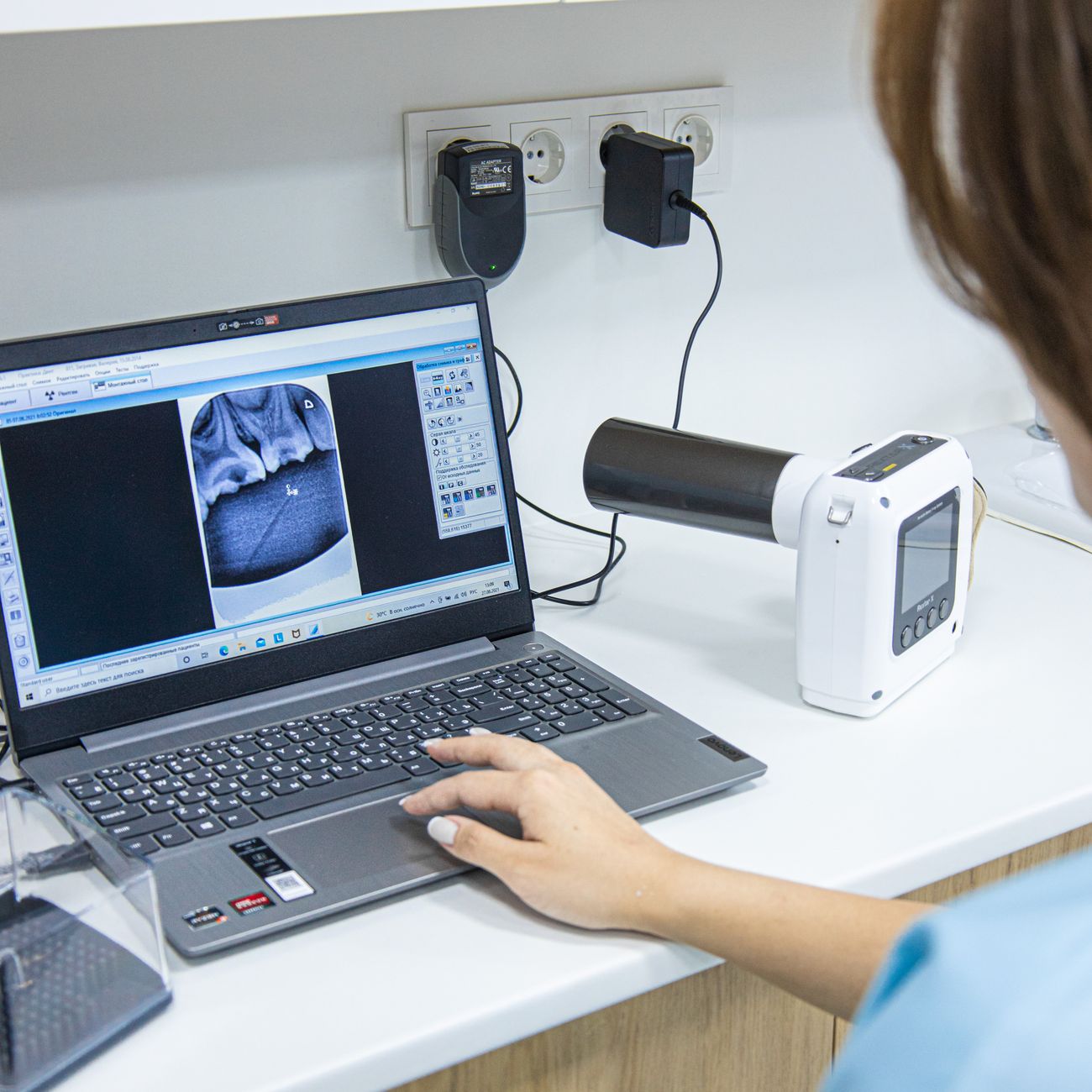
1. Radiography: Radiography is the use of X-rays to create images of the internal structures of the bird. It is a common diagnostic tool in avian medicine and can help identify fractures, foreign bodies, and other abnormalities.
2. Fluoroscopy: Fluoroscopy is a real-time imaging technique that uses X-rays to create moving images of the bird's internal structures. It is useful for evaluating the function of organs such as the heart and gastrointestinal tract.
3. Computed Tomography (CT): CT imaging uses X-rays to create detailed cross-sectional images of the bird's body. It provides more detailed information than traditional radiography and is useful for evaluating complex anatomical structures.
4. Magnetic Resonance Imaging (MRI): MRI uses a magnetic field and radio waves to create detailed images of the bird's internal structures. It is particularly useful for imaging soft tissues such as the brain and spinal cord.
5. Ultrasonography: Ultrasonography uses sound waves to create images of the bird's internal organs. It is a non-invasive imaging technique that is useful for evaluating the liver, kidneys, and reproductive organs.
6. Radiopaque: Radiopaque materials are substances that block X-rays and appear white on radiographs. Examples of radiopaque materials include bones and metal objects.
7. Radiolucent: Radiolucent materials are substances that allow X-rays to pass through and appear dark on radiographs. Examples of radiolucent materials include air and soft tissues.
8. Contrast Agent: A contrast agent is a substance that is used to enhance the visibility of certain structures on radiographs. Contrast agents can be administered orally, intravenously, or intratracheally.
9. Grid: A grid is a device used to improve the quality of radiographic images by reducing scattered radiation. It is placed between the X-ray tube and the bird to improve image contrast.
10. Collimation: Collimation is the process of restricting the X-ray beam to the area of interest. Proper collimation helps reduce radiation exposure to the bird and improves image quality.
11. Digital Radiography: Digital radiography is a modern imaging technique that uses digital sensors to capture X-ray images. It offers several advantages over traditional film-based radiography, including faster image acquisition and the ability to manipulate and store images digitally.
12. PACS (Picture Archiving and Communication System): PACS is a system that allows veterinary professionals to store, retrieve, and view digital radiographic images. It streamlines the image management process and facilitates collaboration between clinicians.
13. DICOM (Digital Imaging and Communications in Medicine): DICOM is a standard protocol used for transmitting and storing medical images, including radiographs. It ensures compatibility between different imaging devices and software systems.
14. Artifact: An artifact is any unintended feature or distortion present in a radiographic image that can affect its interpretation. Common artifacts include grid lines, motion blur, and processing errors.
15. Radiographic Positioning: Proper positioning of the bird is essential for obtaining diagnostic radiographic images. Positioning techniques vary depending on the body part being imaged and may require the use of specialized positioning aids.
16. Ventrodorsal View: A ventrodorsal view is a radiographic projection in which the X-ray beam passes through the bird from ventral to dorsal. It is commonly used to evaluate the chest and abdomen.
17. Lateral View: A lateral view is a radiographic projection in which the X-ray beam passes through the bird from one side to the other. It is useful for evaluating structures such as the wings and legs.
18. Oblique View: An oblique view is a radiographic projection in which the X-ray beam passes through the bird at an angle. It is used to visualize structures that may be obscured on standard views.
19. Myelography: Myelography is a diagnostic procedure that involves injecting a contrast agent into the spinal canal to visualize the spinal cord and nerve roots. It is useful for evaluating spinal cord injuries and disc herniations.
20. Radiographic Interpretation: Radiographic interpretation involves analyzing radiographic images to identify abnormalities and make a diagnosis. It requires knowledge of avian anatomy, pathology, and radiographic principles.
21. Density: Density refers to the degree of blackening on a radiographic image. Bones and metal objects appear white (high density), while air and soft tissues appear dark (low density).
22. Contrast: Contrast refers to the visible difference in density between adjacent structures on a radiographic image. High contrast images have clear distinctions between structures, while low contrast images may appear blurry.
23. Resolution: Resolution is the ability of a radiographic image to show fine details and small structures. High-resolution images have sharp detail, while low-resolution images may lack clarity.
24. Exposure Factors: Exposure factors include factors such as kilovoltage (kV) and milliampere-seconds (mAs) that determine the quality and quantity of X-rays used to create a radiographic image. Proper exposure factors are essential for obtaining diagnostic images.
25. Grid Ratio: Grid ratio refers to the relationship between the height of the lead strips in a grid and the distance between them. Grids with higher ratios are more effective at reducing scattered radiation but may require higher exposure factors.
26. Scatter Radiation: Scatter radiation is the secondary radiation that is produced when X-rays interact with tissues and objects in the bird. It can reduce image quality and increase radiation exposure to the bird and personnel.
27. Radiographic Technique Charts: Technique charts provide guidelines for selecting exposure factors based on the size and anatomy of the bird being imaged. They help ensure consistent image quality and minimize the risk of overexposure.
28. Radiographic Hazards: Radiographic hazards include risks associated with radiation exposure, such as tissue damage, genetic mutations, and increased cancer risk. Proper radiation safety protocols are essential to minimize these risks.
29. Artifacts in Avian Radiology: Artifacts are common in avian radiology and can affect the interpretation of radiographic images. Examples of artifacts include respiratory motion artifacts, grid cutoff, and positioning errors.
30. Avian Radiology Challenges: Avian radiology presents unique challenges due to the small size and delicate anatomy of birds. Factors such as respiratory motion, limited tissue contrast, and positioning difficulties can complicate the imaging process.
31. Avian Radiology Applications: Avian radiology is used in a wide range of clinical scenarios, including the evaluation of fractures, respiratory diseases, gastrointestinal obstructions, and reproductive disorders. It plays a crucial role in diagnosing and monitoring avian patients.
32. Avian Radiology Equipment: Specialized equipment is required for avian radiology, including X-ray machines, digital sensors, collimators, and protective gear. Proper maintenance and calibration of equipment are essential for producing high-quality radiographic images.
33. Avian Radiology Training: Veterinary professionals specializing in avian medicine should undergo training in avian radiology to develop the skills and knowledge necessary to perform and interpret radiographic images effectively. Continuing education is essential to stay updated on advancements in avian imaging technology.
34. Avian Imaging Modalities: In addition to radiography, other imaging modalities such as CT, MRI, and ultrasonography are used in avian medicine to provide complementary information and enhance diagnostic capabilities. Each modality has its strengths and limitations, and the choice of imaging technique depends on the clinical scenario.
35. Avian Radiology Interpretation: Radiographic interpretation in avian medicine requires a thorough understanding of avian anatomy, normal radiographic findings, and common pathological conditions. Veterinarians must be able to differentiate between normal variations and abnormal findings to make accurate diagnoses.
36. Avian Radiology Reporting: Radiology reports should be clear, concise, and comprehensive, providing relevant clinical information, imaging findings, and diagnostic impressions. Effective communication with referring veterinarians and colleagues is essential for optimal patient care.
37. Avian Radiology Quality Control: Quality control measures are essential to ensure the accuracy and reliability of radiographic images in avian medicine. Regular monitoring of equipment performance, image quality, and radiation safety practices is necessary to maintain high standards of care.
38. Avian Radiology Research: Ongoing research in avian radiology aims to improve imaging techniques, develop new diagnostic tools, and enhance our understanding of avian diseases. Collaborative efforts between researchers, clinicians, and imaging specialists are essential to advance the field of avian radiology.
39. Avian Radiology Ethics: Ethical considerations in avian radiology include prioritizing the welfare of the bird, obtaining informed consent for imaging procedures, and respecting client confidentiality. Veterinarians should adhere to professional standards and guidelines to ensure ethical practice.
40. Avian Radiology Future Directions: The future of avian radiology is likely to be shaped by advancements in imaging technology, such as 3D imaging, artificial intelligence, and telemedicine. These innovations have the potential to revolutionize avian diagnostics and improve patient care outcomes.
In conclusion, a solid understanding of key terms and vocabulary in avian radiology and imaging is essential for veterinary professionals working with avian patients. By familiarizing themselves with these concepts, veterinarians can enhance their diagnostic skills, improve patient care, and contribute to the advancement of avian medicine.
Key takeaways
- Understanding key terms and vocabulary in this field is essential for veterinary professionals specializing in avian medicine.
- It is a common diagnostic tool in avian medicine and can help identify fractures, foreign bodies, and other abnormalities.
- Fluoroscopy: Fluoroscopy is a real-time imaging technique that uses X-rays to create moving images of the bird's internal structures.
- It provides more detailed information than traditional radiography and is useful for evaluating complex anatomical structures.
- Magnetic Resonance Imaging (MRI): MRI uses a magnetic field and radio waves to create detailed images of the bird's internal structures.
- It is a non-invasive imaging technique that is useful for evaluating the liver, kidneys, and reproductive organs.
- Radiopaque: Radiopaque materials are substances that block X-rays and appear white on radiographs.